“How are your boobs?”
This is clearly not a usual question. But it’s one a friend asked me, and though I knew why she was asking, it caught me by surprise and made me laugh out loud!
My friend asked me this because the last I had spoken to her, I told her that I was waiting for a biopsy. I had had a mammogram and an ultrasound, and then another mammogram, all within a short period of time because I had found a lump. It wasn’t my first lump. I have what are known as breast density category D (BI-RADS D), indicating “extremely dense” breast tissue, the highest of the four categories.
About 10% of women fall into this category, meaning that the breasts have a high amount of fibrous and glandular tissue. Because this tissue appears white on a mammogram, it makes it tough to distinguish from cancer, which also tends to show up as white. It’s like trying to spot a polar bear in a snow storm. What makes things worse is that dense breasts are also associated with a higher risk of breast cancer because cancers often develop in glandular tissue.
Lucky for me, I don’t have some of the other risk factors that include a family history of breast cancer, genetic mutations associated with higher risk, and prior radiation to the chest area. And, though it’s not always easy to get the imaging care with the frequency needed for denser breasts–I still fit in the “average risk” category with mammograms allowed every 2 years–it is possible to get alternate years of ultrasound, so testing can be done annually. Check your own recommendations here.
Things to Know
Check your breasts. Don’t know how to do a breast exam? Learn how, and do this at least monthly. Here’s a second guide you can download.
In BC, women aged 40-74 may refer themselves to the Breast Screening Program, which means mammograms.
Find out about your breast density.
For those with dense breasts, because of the imaging challenges, you may also be recommended to have a breast ultrasound. Both tests may be recommended because they offer different kinds of information.
The breasts cover a fairly large area, from just below the collarbone (clavicle) to the armpit (axilla) and across to the breastbone (sternum), and they lie over the pectoralis major muscle.
Breasts are made of fat, connective tissue (including fascia and ligaments that support the breast), lymphatic vessels and nodes, glands (15-25 groups of glands called lobules that make milk in each breast), and ducts (tubes that carry milk from the lobules to the nipple).
Female breasts are active organs because they contain active glandular tissue and are hormonally responsive.
Speaking of hormones, you may already know this from your own personal experience, but breast tissue changes with hormonal shifts. Swelling, changes in density, cyst size, tenderness, and other sensations can often be felt particularly in the days before menstruation. But they also change with puberty, during perimenopause and menopause, and in relationship to pregnancy and breastfeeding.
Male breasts are obviously normally less developed, so less than 1% of breast cancers occur in men. However, if you notice a lump, a discharge or bleeding from the nipple, or other changes in colour, skin, or discomfort, talk to your physician.
What Did I Learn?
First and foremost, I learned that you really do have to advocate for yourself. Sometimes you have to be politely insistent. The last time I had a lump checked, I had been told about my D breasts (no, not the cup size! haha!), and I was told at the imaging centre that I would need to get alternating years of mammogram and ultrasound. It wasn’t made clear to me how that would happen, and I had thought that they would contact me.
When I started to be bothered by another lump (it was particularly sore), I realized it had been more than a year since my last imaging, so I went into their office to check for a booking (they often don’t answer their phones!). They told me it was too early and I’d have to wait another 6 months. I told them I had been previously told I should be approved for some type of imaging annually. They said they checked my file and I’d have to wait. I insisted that I had a lump of concern. They told me it would be another few months, as they were heavily booked. I said that seemed too late. They found me an ultrasound booking and told me to get a note from my MD and bring that in with me at that appointment so I would not be charged for a late cancellation, as without it, they would not do the imaging. I’m lucky to have a GP, so I did just that.
At the ultrasound, the tech told me it was good I had pushed, as she was the one who had last noted the need for more regular imaging. She found 2 lumps of concern. So, I learned I was right in my memory of the information I had been given, despite being told at the office that I was wrong. Trust yourself. But it’s probably also a good idea to write down what you’re told when you’re told, so you can be sure. 🙂
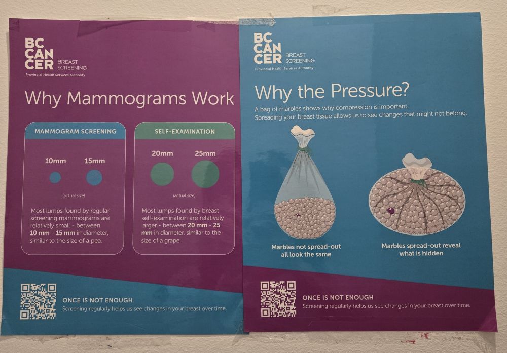
While no one loves getting a mammogram because they are uncomfortable, they needn’t be horribly painful! Usually, when I’ve had mammograms, they’ve been moderately uncomfortable. But one of my mammograms (4 images taken, so 4 times I had to bear through it) was so painful that I couldn’t breathe normally. I told her so as she set me up for the first image. She said it had to be tight. I didn’t speak up further. Later, during my biopsy, I asked the physician what level of pain is reasonable. She said that while flattening the breasts as best as possible improves the image, you should certainly be able to breathe. It is uncomfortable, but should not be gritting teeth, holding breath, squealing pain. Know your pain level and speak up if it’s too much.
Don’t let the fear of discomfort stop you from getting a mammogram. When done properly, it’s really quite bearable. And, if it goes beyond, tell them that the pressure is painful.
The last time I had a lump, it was found to be just a large, fluid-filled cyst. I had asked if it could be drained, as it was painful. I was told then, that there was no point, as it would simply return. I found out this time that if the lump is large, painful, or causing discomfort, it can absolutely be drained. They use a fine-needle that they direct under the visuals of an ultrasound to draw out the fluid.
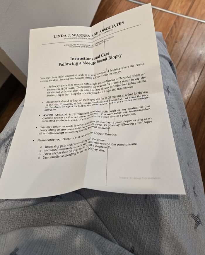
The biopsy was easy. If you have to get one, it’s not bad. But you may be sent home with a bag of frozen peas to tuck into your bra, as you’ll need to ice the area on and off for the rest of the day. If you plan to return to your workplace after, that might make things a bit awkward!
You’ll also have a little titanium marker left inside where the biopsy is done to mark the site for future imaging or for surgery. If you feel a slight buzzing sensation after (as I did), it’s just a slightly irritated nerve, and it should go away. For me, it stopped after a couple of weeks, though I could not sleep on that side for longer.

Why Share This Information?
I don’t normally get this personal in a blog. But I think it’s important to talk about a part of our anatomy that we normally don’t discuss. How many of you knew all these things about breasts?
I was going to try to write this blog in March, closer to International Women’s Day, but the month flew by! It’s timely year-round, but perhaps even more relevant in April, during Cancer Awareness Month. Because prevention and detection are important for better outcomes.
In Traditional Chinese Medicine, there are several acupuncture channels that travel through the chest and breasts: Lungs, Pericardium, Spleen, Stomach, Gallbladder, Kidneys, and Ren. But it’s not just the visible points that have impact on this tissue. So, while you should do regular breast self-exams and get imaging, you can also work on breast health through all the usual wellness care therapies of TCM.